


Frequently bought together
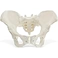
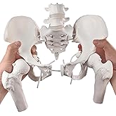
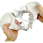
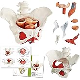
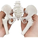
This item: MonMed Life Size Female Pelvis Model, Hip Model – Female Anatomy Model, Hip Bone Pelvic Model Female Anatomical Model
$43.53
Get it by Saturday, Nov 22
Only 9 left in stock.
+
$52.50
Get it by Thursday, Nov 27
Only 2 left in stock.
Total price:��$00
To see our price, add these items to your cart.
Choose items to buy together.
Customers also viewed these products
Page 1 of 1 Start again
- Evotech Flexible Female Pelvis Model On Elastic, Life Size Female Pelvic Skeleton Model W/Bungee, Anatomy Medical Model for Science Education, Midwife in Obstetrics, Gynecology & Patent CommunicationFREE Shipping by �鶹��Get it by Thursday, Nov 27Only 6 left in stock.
- Benilev Female Pelvis Model with Pelvic Floor Muscles Perineal Model Female Anatomy Model 1:1 Life Size Human Skeleton Medical Science Educational Equipment Anatomy MedicineFREE Shipping by �鶹��Get it by Friday, Nov 21


Brand in this category on �鶹��
Sponsored

Brands in this category on �鶹��
Sponsored


